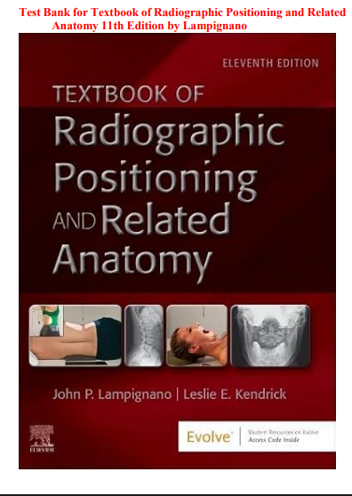


Radiography students learn two skills at once. They must know the anatomy well enough to name every structure on an image, and they must know how to place a patient, the central ray, and the image receptor so those structures show up clearly. Exams test both, often in a single question. A question may describe a poor image and ask what went wrong, or name an injury and ask which projection shows it best. This test bank was built to give you practice with that kind of thinking. It follows the chapter order of Bontrager’s Textbook of Radiographic Positioning and Related Anatomy, 11th Edition, by John Lampignano and Leslie E. Kendrick, so you can study each body region in the same sequence as your course.
If you are in a radiologic technology program, preparing for a positioning competency, or reviewing for the ARRT exam, this resource gives you steady practice with the questions you will meet most often.
Topics Covered
The question sets follow the textbook and include:
- Terminology, patient care, and basic positioning language
- Radiation protection, including time, distance, shielding, and ALARA
- Digital imaging, exposure factors, and image quality
- Chest and thoracic viscera, including routine and decubitus projections
- Abdomen and the digestive system, including contrast studies
- Urinary system studies and contrast safety
- Upper extremity: fingers, hand, wrist, forearm, and elbow
- Shoulder girdle and humerus
- Lower extremity: toes, foot, ankle, leg, knee, and femur
- Pelvis and hip
- Vertebral column, from cervical through sacrum and coccyx
- Bony thorax: ribs and sternum
- Skull, facial bones, and sinuses
- Trauma, mobile, and surgical radiography
- Pediatric and older adult imaging
- Introductions to CT, MRI, nuclear medicine, and other modalities
Questions are a mix of multiple-choice, matching, and short scenario items, which are the formats positioning courses and registry-style exams use most.
Why Practice Questions Help in This Course
Positioning has a lot of small details that look alike. A few degrees of rotation, one angle of the central ray, or one different landmark can change which structures appear and which are hidden. Practicing with questions helps you:
- Connect each projection with the structure it is meant to show
- Recall central ray angles, entry points, and patient positions without guessing
- Find the cause of a poor image, such as rotation, motion, or wrong centering
- Apply radiation safety rules automatically in routine and portable work
- Get used to the way exam questions are worded
Many students say they understand a position while reading, then lose the details when a question changes one factor. Regular practice by chapter makes those details stick.
What Makes This Test Bank Different
Every question includes a full rationale. You will see why the correct answer is right, what the image or position would look like with a wrong choice, and why each incorrect option does not fit. Incorrect options in positioning questions are usually real projections or real angles used for a different purpose, so learning why they are wrong helps you remember when each one is right.
Questions are grouped by chapter and body region, so you can review one area, such as the upper extremity, before a lab check-off, or work through several regions before a comprehensive exam. Use it beside your reading, before clinical, or as a structured review before the registry exam.
Sample Questions
Question 1
Why is a PA chest radiograph taken at a source-to-image distance of 72 inches (183 cm) instead of a shorter distance?
A. To reduce magnification of the heart and give sharper detail of the lungs
B. To increase the radiation dose to the patient for better contrast
C. To make the exposure time longer so motion is easier to control
D. To make the heart appear larger so it is easier to measure
Correct Answer: A
Rationale: A longer source-to-image distance makes the x-ray beam less divergent, which reduces magnification and improves sharpness. This matters in chest imaging because the heart is a standard size reference, and magnification would make it look larger than it is. Increasing the dose is not a goal, and a longer distance actually requires more exposure to compensate. A longer distance does not make a long exposure desirable, since short exposure times are preferred to limit motion. Magnifying the heart would make heart size measurements unreliable.
Question 2
On an oblique lumbar spine image, which part of the “Scottie dog” represents the pars interarticularis?
A. The eye
B. The nose
C. The ear
D. The neck
Correct Answer: D
Rationale: On a properly positioned oblique lumbar projection, the posterior elements form an image that looks like a Scottie dog. The pars interarticularis forms the neck, which is where a defect called spondylolysis shows up as a “collar” on the dog. The pedicle forms the eye, the transverse process forms the nose, and the superior articular process forms the ear. Knowing which structure each part represents helps you judge whether the obliquity is correct and where a defect would appear.
Question 3
A technologist is performing a portable chest exam at the bedside and wants to reduce occupational exposure. Which action is most effective?
A. Standing as far from the x-ray tube as the exposure cord allows, ideally at least 6 feet (2 m)
B. Standing directly behind the patient’s bed rail
C. Holding the image receptor in place with a bare hand
D. Using a higher mAs to shorten the exposure
Correct Answer: A
Rationale: Distance is one of the three basic protection principles, along with time and shielding. Because exposure from scatter drops quickly as you move away from the source, standing at least 6 feet away and wearing a lead apron if within range greatly reduces dose. A bed rail does not shield the technologist from scatter. Holding the image receptor with a bare hand puts the hand in or near the beam and is unsafe. Using a higher mAs does not shorten the exposure and would increase the amount of scatter produced.
Question 4
Which projection of the shoulder places the glenoid fossa in profile so the glenohumeral joint space appears open?
A. AP oblique (Grashey method) with the patient rotated about 35 to 45 degrees toward the affected side
B. Transthoracic lateral
C. Tangential (Fisk) projection of the bicipital groove
D. AP clavicle projection with a cephalic angle
Correct Answer: A
Rationale: In the Grashey method, the patient is rotated toward the affected shoulder so the scapula lies flat against the image receptor. The glenoid fossa is then seen in profile, which opens the joint space and shows the humeral head clearly. This is helpful for evaluating dislocations and joint arthritis. A transthoracic lateral looks at the proximal humerus from the side. A tangential projection is meant to show the bicipital groove, and an AP clavicle with an angle is used for the clavicle, not the glenohumeral joint space.
Question 5
A patient has snuffbox tenderness after a fall on an outstretched hand, and a scaphoid fracture is suspected. Which projection best demonstrates the scaphoid?
A. PA wrist with ulnar deviation
B. Lateral wrist in neutral position
C. AP forearm
D. PA wrist with radial deviation
Correct Answer: A
Rationale: Ulnar deviation moves the scaphoid away from other carpal bones and straightens it, so it is shown at its full length, which helps reveal subtle fracture lines. Snuffbox tenderness is a classic clinical sign of a scaphoid injury. A neutral lateral wrist overlaps carpal bones, making the scaphoid hard to see. An AP forearm is used for forearm bones and does not show the carpal bones well. Radial deviation foreshortens the scaphoid, which makes a fracture harder to find.
Frequently Asked Questions
Is this the same as the publisher’s official test bank?
No. This is an independently written study resource built to help students review the topics in the 11th edition of Bontrager’s Textbook of Radiographic Positioning and Related Anatomy. It is a self-study companion and is not distributed by the publisher.
Will it help me prepare for the ARRT exam?
The questions focus on positioning, anatomy, image evaluation, and radiation protection, which are heavily tested areas on the registry exam. It works best as practice alongside your textbook and clinical experience.
Does it cover every chapter?
Yes. Questions are organized by chapter and body region, following the order of the 11th edition from terminology and radiation protection through each body region and special modalities.
What kinds of questions are included?
Mostly multiple-choice, with some matching and short scenario questions.
Are there explanations or only correct letters?
Every question includes a full rationale explaining why the correct answer is right and why the other options do not fit.
Who is it best for?
Students in radiologic technology programs, and anyone reviewing positioning and anatomy before a course exam, competency check, or registry exam.
How should I use it?
Read the chapter first, then answer the questions without checking the answers. Study the rationales afterward, especially for questions you missed, since understanding why a position works is more useful than memorizing a letter.
Can it replace the textbook or lab practice?
No. It works best alongside the textbook and hands-on practice. The book teaches the positions, and the test bank helps you recall and apply the details.
Is it matched to the 11th edition?
Yes. It follows the topics, terminology, and chapter structure of the 11th edition.
Is it useful the night before an exam?
Yes. Because it is organized by chapter and body region, you can quickly review a weak area, such as the shoulder or lumbar spine, without going back through the whole book.




Zack Evans –
Excellent nursing test bank resources
Joel Terry –
Very helpful for exam preparation
Ann –
Helpful questions that build confidence.
Paulinetops1 –
Excellent study aid for nursing exams.
Swagridi –
Easy to use and very informative
Nichola Sabraico –
Excellent nursing questions and answers.
Harry Jans –
Very helpful for daily nursing studies